|
 |
Photo: Supplied
08 May 2013 |
No, a glass of champagne will not cure cancer....
…But they have more in common than you might think.
Researchers from the Departments of Microbial Biochemical and Food Biotechnology, Physics and the Centre for Microscopy at the University of the Free State in South Africa were recently exploring the properties of yeast cells in wine and food to find out more of how yeast was able to manufacture the gas that caused bread to rise, champagne to fizz and traditional beer to foam. And the discovery they made is a breakthrough that may have enormous implications for the treatment of diseases in humans.
The team discovered that they could slice open cells with argon gas particles, and look inside. They were surprised to find a maze of tiny passages like gas chambers that allowed each cell to ‘breathe.’ It is this tiny set of ‘lungs’ that puts the bubbles in your bubbly and the bounce in your bread.
But it was the technique that the researchers used to open up the cells that caught the attention of the scientists at the Mayo Clinic (Tumor Angiogenesis and Vascular Biology Research Centre) in the US.
Using this technology, they ultimately aim to peer inside cells taken from a cancer patient to see how treatment was progressing. In this way they would be able to assist the Mayo team to target treatments more effectively, reduce dosages in order to make treatment gentler on the patient, and have an accurate view of how the cancer was being eliminated.
“Yes, we are working with the Mayo Clinic,” said Profes Lodewyk Kock from the Microbial, Biochemical and Food Biotechnology Department at the UFS.
“This technique we developed has enormous potential for cell research, whether it is for cancer treatment or any other investigation into the working of cells. Through nanotechnology, and our own invention called Auger-architectomics, we are able to see where no-one has been able to see before.”
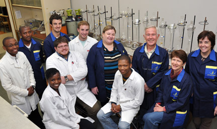
The team of Prof Kock including Dr Chantel Swart, Kumisho Dithebe, Prof Hendrik Swart (Physics, UFS) and Prof Pieter van Wyk (Centre for Microscopy, UFS) unlocked the ‘missing link’ that explains the existence of bubbles inside yeasts, and incidentally have created a possible technique for tracking drug and chemotherapy treatment in human cells.
Their work has been published recently in FEMS Yeast Research, the leading international journal on yeast research. In addition, their discovery has been selected for display on the cover page of all 2013 issues of this journal.
One can most certainly raise a glass of champagne to celebrate that!
There are links for video lectures on the technique used and findings on the Internet at:
1. http://vimeo.com/63643628 (Comic version for school kids)
2. http://vimeo.com/61521401 (Detailed version for fellow scientists)