Latest News Archive
Please select Category, Year, and then Month to display items
07 January 2018
Photo Charl Devenish
 Dr Anneke van der Spoel van Dijk is invested in contributing
to the global effort of stopping TB by 2035.
Dr Anneke van der Spoel van Dijk is invested in contributing
to the global effort of stopping TB by 2035.
The work of Dr Anneke van der Spoel van Dijk investigates the spread of TB in the Free State population using techniques such as next generation sequencing, spoligotyping and MIRU-VNTR typing. Dr Van der Spoel van Dijk, a senior medical scientist in the Department of Medical Microbiology at the University of the Free State (UFS) also looks at drug resistance in her research. This work informs decisions about how best to treat patients with multidrug-resistant TB (MDR-TB).
She employs rapid molecular techniques to track one of Africa’s most serious diseases, tuberculosis (TB).
Drug resistance
Scientists assist the National Health Laboratory Service and Department of Health in trying to refine the diagnostic tools to identify these cases earlier. Dr Van der Spoel van Dijk explains: “Until recently, it took up to two years to fine-tune treatment decisions for patients with MDR-TB. Patients get a cocktail of anti-TB drugs, but it takes time to find the right combination. Re-infection and relapse (patients stopping treatment for several reasons) add to the diagnostic and treatment management challenges.
Enormous impact
“Now doctors can reduce the time needed for diagnostic certainty to about seven days, while new drugs allow reduction of treatment from more than 18 to nine months. This can have an enormous impact on the life of many patients.”
Dr Van der Spoel van Dijk’s work forms part of research in the faculty looking at resistance development in TB strains. She is currently also doing her doctoral thesis on the differences and incidence of MDR-TB among adolescents versus adults. Dr Van der Spoel van Dijk says: “It is a complicated picture, but we hope to unravel it to support better diagnostic tools and patient care.”
As part of the National Health Laboratory Service, her department is playing an important role in TB diagnostics and the training of scientists and future pathologists. “Our work is contributing to the global vision to stop TB by 2035,” Dr Van der Spoel van Dijk says.
Mathematical methods used to detect and classify breast cancer masses
2016-08-10
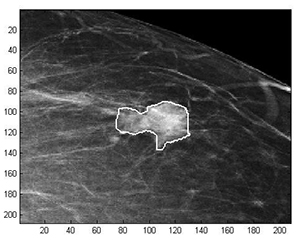 Examples of Acho’s breast mass
segmentation identification
Breast cancer is the leading cause of female mortality in developing countries. According to the World Health Organization (WHO), the low survival rates in developing countries are mainly due to the lack of early detection and adequate diagnosis programs.
Seeing the picture more clearly
Susan Acho from the University of the Free State’s Department of Medical Physics, breast cancer research focuses on using mathematical methods to delineate and classify breast masses. Advancements in medical research have led to remarkable progress in breast cancer detection, however, according to Acho, the methods of diagnosis currently available commercially, lack a detailed finesse in accurately identifying the boundaries of breast mass lesions.
Inspiration drawn from pioneer
Drawing inspiration from the Mammography Computer Aided Diagnosis Development and Implementation (CAADI) project, which was the brainchild Prof William Rae, Head of the department of Medical Physics, Acho’s MMedSc thesis titled ‘Segmentation and Quantitative Characterisation of Breast Masses Imaged using Digital Mammography’ investigates classical segmentation algorithms, texture features and classification of breast masses in mammography. It is a rare research topic in South Africa.
Characterisation of breast masses, involves delineating and analysing the breast mass region on a mammogram in order to determine its shape, margin and texture composition. Computer-aided diagnosis (CAD) program detects the outline of the mass lesion, and uses this information together with its texture features to determine the clinical traits of the mass. CAD programs mark suspicious areas for second look or areas on a mammogram that the radiologist might have overlooked. It can act as an independent double reader of a mammogram in institutions where there is a shortage of trained mammogram readers.
Light at the end of the tunnel
Breast cancer is one of the most common malignancies among females in South Africa. “The challenge is being able to apply these mathematical methods in the medical field to help find solutions to specific medical problems, and that’s what I hope my research will do,” she says.
By using mathematics, physics and digital imaging to understand breast masses on mammograms, her research bridges the gap between these fields to provide algorithms which are applicable in medical image interpretation.