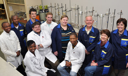
Latest News Archive

Please select Category, Year, and then Month to display items
Previous Archive
26 September 2018
The Cardiac Simulation lab in action

There’s an electric atmosphere in the operating theatre of the Faculty of Health Sciences, as Dr Taha Gwila and his team focus with intense concentration on the fleshy exposed heart beating rhythmically in the opened chest of the patient lying in front of them. The enormous demands of open-heart surgery are evident to everyone looking on. But there’s a catch. 

The patient is faceless and rubberised. The red liquid flowing in the pipes that network from the body is not blood. And the pulsating heart was beating in the body of a pig not too long ago. 

Cutting edge technology
 
This Cardiac Simulation laboratory supplied by Medtronic is the newest addition to the School of Biomedical Sciences’ clinical simulation and skills unit. 

“There’s nothing like this in Africa, and only a few in the world,” says a beaming Prof Francis Smit, Head of Cardiothoracic Surgery at the Faculty of Health Sciences.
He explains that this new cutting edge medical technology will revolutionise the way cardiac surgeons and other health professionals are trained and assessed.

Practicing specific procedures

The simulation facilities give students with various levels of competency the opportunity to practice specific procedures in their own time and at their own pace.
“Traditionally training followed the apprentice model, where surgeons started with simple tasks and worked their way up. They assisted senior personnel and their exposure to procedures depended on the conditions presented by the patients before them,” explains Prof Smit.

The simulation technology now enables them to repeatedly practise a certain procedure without any risk to a patient. A sophisticated electronic grading system gives detailed feedback after each session, so they know in which areas to improve.  

Simulated emergencies

The system also allows trainers to create a medical emergency that the trainees then have to deal with.

“Assisting senior surgeons with high levels of competency means that in the past, trainees would often never get the chance to experience these kinds of complications during operating procedures. Now we give them a chance to build that confidence so they’ll be able to handle different situations.”  

Training hub for Africa
 

The UFS cardiothoracic programme is being designed to become a training hub for the whole of Southern Africa, combining distance learning with an on-site high-fidelity simulation and assessment centre.

“This is 100% real!” says an excited Dr Gwila after successfully completing his first simulation session. “As a Senior Registrar at the Cardiothoracic Department I’ve done similar procedures on real patients and there’s really no difference at all. Every registrar should do this before ever touching a real body.”

News Archive

Champagne and cancer have more in common than you might think
2013-05-08


Photo: Supplied
08 May 2013

No, a glass of champagne will not cure cancer....

…But they have more in common than you might think.

Researchers from the Departments of Microbial Biochemical and Food Biotechnology, Physics and the Centre for Microscopy at the University of the Free State in South Africa were recently exploring the properties of yeast cells in wine and food to find out more of how yeast was able to manufacture the gas that caused bread to rise, champagne to fizz and traditional beer to foam. And the discovery they made is a breakthrough that may have enormous implications for the treatment of diseases in humans.

The team discovered that they could slice open cells with argon gas particles, and look inside. They were surprised to find a maze of tiny passages like gas chambers that allowed each cell to ‘breathe.’ It is this tiny set of ‘lungs’ that puts the bubbles in your bubbly and the bounce in your bread.

But it was the technique that the researchers used to open up the cells that caught the attention of the scientists at the Mayo Clinic (Tumor Angiogenesis and Vascular Biology Research Centre) in the US.

Using this technology, they ultimately aim to peer inside cells taken from a cancer patient to see how treatment was progressing. In this way they would be able to assist the Mayo team to target treatments more effectively, reduce dosages in order to make treatment gentler on the patient, and have an accurate view of how the cancer was being eliminated.

“Yes, we are working with the Mayo Clinic,” said Profes Lodewyk Kock from the Microbial, Biochemical and Food Biotechnology Department at the UFS.

“This technique we developed has enormous potential for cell research, whether it is for cancer treatment or any other investigation into the working of cells. Through nanotechnology, and our own invention called Auger-architectomics, we are able to see where no-one has been able to see before.”

The team of Prof Kock including Dr Chantel Swart, Kumisho Dithebe, Prof Hendrik Swart (Physics, UFS) and Prof Pieter van Wyk (Centre for Microscopy, UFS) unlocked the ‘missing link’ that explains the existence of bubbles inside yeasts, and incidentally have created a possible technique for tracking drug and chemotherapy treatment in human cells.

Their work has been published recently in FEMS Yeast Research, the leading international journal on yeast research. In addition, their discovery has been selected for display on the cover page of all 2013 issues of this journal.

One can most certainly raise a glass of champagne to celebrate that!

There are links for video lectures on the technique used and findings on the Internet at:

1. http://vimeo.com/63643628 (Comic version for school kids)

2. http://vimeo.com/61521401 (Detailed version for fellow scientists)

We use cookies to make interactions with our websites and services easy and meaningful. To better understand how they are used, read more about the UFS cookie policy. By continuing to use this site you are giving us your consent to do this.

Accept