Latest News Archive

Please select Category, Year, and then Month to display items
Previous Archive
21 November 2019 | Story Nonsindiso Qwabe | Photo Charl Devenish
Ultrasound read more
Checking out some features of the Samsung ultrasound system are, from the left: SSEM Mthembu Medical's Chase Hutchinson and Jannie Coetzee; Head of Anaesthesiology, Dr Edwin Turton; and Head of Undergraduate Training in Anaesthesiology, Prof Lomby Odendaal.

Medical students in the Faculty of Health Sciences at the UFS will now be able to learn how to perform procedures such as the precise location of a vein for intravenous lines and for diagnostic procedures such as detecting abnormalities in pregnancies, identifying gallstones, and diagnosing trauma-related injuries with ease.  This will be made possible by the placement of a one-of-a-kind ultrasound machine – putting them on par with cutting-edge global medical technology.

A first ever in the medical curriculum of undergraduate students at the UFS

The state-of-the-art, compact HS70A Samsung ultrasound system to the value of R1,4 million was unveiled in the Faculty of Health Sciences’ Clinical Simulation and Skills Unit on 19 November. A first ever in the medical curriculum of undergraduate students at the UFS, it is set to revolutionise the delivery of health-care education in the faculty, said Prof Lomby Odendaal, Teaching and Learning Coordinator for undergraduate anaesthesiology training in the Department of Anaesthesiology.

The ultrasound system was donated by SSEM Mthembu Medical and Samsung Korea.
Prof Odendaal said for the first time in the history of the undergraduate MB ChB curriculum, the ultrasound will be available to medical students from their third year. Students have never had the opportunity to be trained in using ultrasound this early in their careers.

Improved clinical training experience of students

Ultrasound is a diagnostic medical tool that uses sound waves to produce images of internal structures of the body. Prof Odendaal said ultrasound is important to determine pathology and diseases in the body and to provide point-of-care ultrasound. Having the ultrasound in the unit will transform the clinical training experience of students, training them to provide better treatment and medical care, even in constrained environments, to improve patient care.

“There is almost no structure in the body that cannot be examined using ultrasound. It makes the delivery of healthcare more effective. If you make a better diagnosis, the treatment and care will be much better. Ultrasound is so important lately that if you don’t do it, you will be left behind. That’s why we decided to bring this to the students. We can’t miss out on teaching our students about ultrasound, because we want them to be familiar with it by the time they finish their medical degree, so that, even if they go to smaller hospitals, they will be able to spread diagnostic care to the periphery,” Prof Odendaal said.

Streamlined workflow for patient care

“The cutting-edge technology and rich image quality of the ultrasound will deliver top-notch diagnoses to suit the diverse departments within the faculty,” said Chase Hutchinson, National Product Manager at SSEM Mthembu Medical. It comes with various pre-set models to cater for different needs and applications, allowing streamlined workflow for higher efficiency and patient care.

According to Prof Mathys Labuschagne, the Head of the Clinical Simulation and Skills Unit, ultrasound training will improve the quality of doctors graduating in the faculty. “We are really excited about this. You can diagnose many conditions using ultrasound and deliver point-of-care ultrasound; this will become a natural part of students’ training and clinical practice in future.”

News Archive

Nanotechnology breakthrough at UFS
2010-08-19

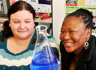
 Ph.D students, Chantel Swart and Ntsoaki Leeuw


Scientists at the University of the Free State (UFS) made an important breakthrough in the use of nanotechnology in medical and biological research. The UFS team’s research has been accepted for publication by the internationally accredited Canadian Journal of Microbiology.

The UFS study dissected yeast cells exposed to over-used cooking oil by peeling microscopically thin layers off the yeast cells through the use of nanotechnology.

The yeast cells were enlarged thousands of times to study what was going on inside the cells, whilst at the same time establishing the chemical elements the cells are composed of. This was done by making microscopically small surgical incisions into the cell walls.

This groundbreaking research opens up a host of new uses for nanotechnology, as it was the first study ever in which biological cells were surgically manipulated and at the same time elemental analysis performed through nanotechnology. According to Prof. Lodewyk Kock, head of the Division Lipid Biotechnology at the UFS, the study has far reaching implications for biological and medical research.

The research was the result of collaboration between the Department of Microbial, Biochemical and Food Biotechnology, the Department of Physics (under the leadership of Prof. Hendrik Swart) and the Centre for Microscopy (under the leadership of Prof.Pieter van Wyk).

Two Ph.D. students, Chantel Swart and Ntsoaki Leeuw, overseen by professors Kock and Van Wyk, managed to successfully prepare yeast that was exposed to over-used cooking oil (used for deep frying of food) for this first ever method of nanotechnological research.

According to Prof. Kock, a single yeast cell is approximately 5 micrometres long. “A micrometre is one millionth of a metre – in laymen’s terms, even less than the diameter of a single hair – and completely invisible to the human eye.”

Through the use of nanotechnology, the chemical composition of the surface of the yeast cells could be established by making a surgical incision into the surface. The cells could be peeled off in layers of approximately three (3) nanometres at a time to establish the effect of the oil on the yeast cell’s composition. A nanometre is one thousandth of a micrometre.

Each cell was enlarged by between 40 000 and 50 000 times. This was done by using the Department of Physics’ PHI700 Scanning Auger Nanoprobe linked to a Scanning Electron Microscope and Argon-etching. Under the guidance of Prof. Swart, Mss. Swart en Leeuw could dissect the surfaces of yeast cells exposed to over-used cooking oil. 

The study noted wart like outgrowths - some only a few nanometres in diameter – on the cell surfaces. Research concluded that these outgrowths were caused by the oil. The exposure to the oil also drastically hampered the growth of the yeast cells. (See figure 1)  

Researchers worldwide have warned about the over-usage of cooking oil for deep frying of food, as it can be linked to the cause of diseases like cancer. The over-usage of cooking oil in the preparation of food is therefore strictly regulated by laws worldwide.

The UFS-research doesn’t only show that over-used cooking oil is harmful to micro-organisms like yeast, but also suggests how nanotechnology can be used in biological and medical research on, amongst others, cancer cells.
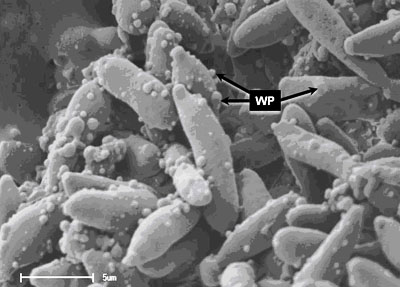
 

Figure 1. Yeast cells exposed to over-used cooking oil. Wart like protuberances/ outgrowths (WP) is clearly visible on the surfaces of the elongated yeast cells. With the use of nanotechnology, it is possible to peel off the warts – some with a diameter of only a few nanometres – in layers only a few nanometres thick. At the same time, the 3D-structure of the warts as well as its chemical composition can be established.  

Media Release
Issued by: Mangaliso Radebe
Assistant Director: Media Liaison
Tel: 051 401 2828
Cell: 078 460 3320
E-mail: radebemt@ufs.ac.za  
18 August 2010
 

We use cookies to make interactions with our websites and services easy and meaningful. To better understand how they are used, read more about the UFS cookie policy. By continuing to use this site you are giving us your consent to do this.

Accept