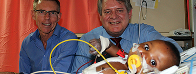
Photo: René-Jean van der Berg |
A team from the University of the Free State School for Medicine work daily unremittingly to save the lives of young children who have been born with heart defects by carrying out highly specialised interventions and operations on them. These operations, which are nowadays performed more and more frequently by cardiologists from the UFS School of Medicine, place the UFS on a similar footing to world-class cardiology and cardio-thoracic units.
One of the children is seven-month-old Montsheng Ketso who recently underwent a major heart operation to keep the left ventricle of her heart going artificially.
Montsheng was born with a rare, serious defect of the coronary artery, preventing the left ventricle from receiving enough blood to pump to the rest of the body.
This means that the heart muscle can suffer damage because these children essentially experience a heart attack at a very young age.
In a healthy heart, the left ventricle receives oxygenated blood from the left atrium. Then the left ventricle pumps this oxygen-rich blood to the aorta whence it flows to the rest of the body. The heart muscle normally receives blood supply from the oxygenated aorta blood, which in this case cannot happen.
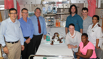
Photo: René-Jean van der Berg |
“She was very ill. I thought my baby was going to die,” says Mrs Bonizele Ketso, Montsheng’s mother.
She says that Montsheng became sick early in February, and she thought initially it was a tight chest or a cold. After a doctor examined and treated her baby, Montsheng still remained constantly ill, so the doctor referred her to Prof Stephen Brown, paediatric cardiologist at the UFS and attached to Universitas Hospital.
Here, Prof Brown immediately got his skilled team together as quickly as possible to diagnose the condition in order to operate on Montsheng.
During the operation, the blood flow was restored, but since Montsheng’s heart muscle was seriously damaged, the heart was unable to contract at the end of the operation. Then she was coupled to a heart-lung machine to allow the heart to rest and give the heart muscle chance to recover. The entire team of technologists and the dedicated anaesthetist, Dr Edwin Turton, kept a vigil day and night for several days.
Prof Francis Smit, chief specialist at the UFS Department of Cardiothoracic Surgery, explains that without this operation Montsheng would not have been able to celebrate her first birthday.
“After the surgery, these children can reach adulthood without further operations. Within two to three months after the operation, she will have a normal active life, although for about six months she will still use medication. Thereafter, she will be tiptop and shortly learn to crawl and walk.”
Mrs Ketso is looking forward enormously to seeing her daughter stand up and take her first steps. A dream which she thought would never come true.
“Write there that I really love these doctors.”