Latest News Archive

Please select Category, Year, and then Month to display items
Previous Archive
09 December 2019 | Story Nonsindiso Qwabe | Photo Barend Nagel
Victoria Read more
Victoria the mannequin has become a familiar face in Nursing classrooms

She has an uncanny ability to move her eyes towards the sound of voices in a room, her voice shrills and squeals when she's in pain, she throws in a Spanish word or two, and she releases bodily fluids just like a real human would. 

These are just some of the quirky characteristics that make up the new R1,76 million-rand birthing mannequin in the School of Nursing's Simulation Unit, who goes by the name Victoria. She weighs more than 80 kg and is almost 1,7 metres high. With features such as real eyelashes, eyebrows, and hair, you can't help but do a double take when you lay eyes on her. 

Students getting practical experience

While the unit has other mannequins used for training Nursing students in each year or their study, Simulation Coordinator from the School of Nursing in the Faculty of Health Sciences, Cecile Fourie, said Victoria was a major upgrade for the school because of her versatility. Victoria would further enhance the school's quality of teaching by training students in their final year of undergraduate studies as well as those pursuing their postgraduate studies, about the ins and outs of pregnancy and other female morbidities, Fourie said. 

"We try to make our scenarios as real and authentic as possible and we've seen how much our students have grown. Introducing Victoria to our pre- and postgraduate students will prepare them to be competent in clinical practice." 

Meet Victoria

So, what exactly can Victoria do? 

Fourie said while the other mannequins were made with screws that made them look robot-like, Victoria was made with silicone and given a clean finish to make her appear life-like. The other mannequins can only blink, but Victoria's eyes move around, she can speak Spanish and French, and she comes with five tummies for different medical scenarios. She has a normal tummy which acts as a closure, an operable tummy for caesarean delivery, a tummy that allows a breeched baby to be twisted and turned from the outside just before delivery, a contraction tummy used for normal vaginal delivery – which also allows Victoria to push, bleed, urinate, and release mineral oil which acts as amniotic fluid. The fifth tummy is postpartum haemorrhage, a condition that is common among South African mothers after delivery. 

"It’s so good that we get to train our students in such real, lifelike circumstances. Through Victoria we're going to try and do our part in lessening maternal deaths, which are so prominent in our country. She can act out all the abnormalities that take place in a real delivery, and she can also have other medical conditions such as a heart attack," Fourie said.


News Archive

Collaboration between UFS and Mayo Clinic to revolutionise cancer treatment
2014-06-27


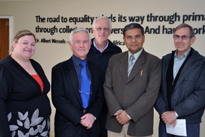
Attending the lecture were, from the left: Dr Chantel Swart, Prof Lodewyk Kock, Prof Debabrata Mukhopadhyay, Prof James du Preez; back: Prof Pieter van Wyk.
Dr Swart, Profs Kock and Du Preez are from the Department of Microbial, Biochemical and Food Biotechnology. Prof Mukhopadhyay is from the Mayo Clinic (US) and Prof Van Wyk is from the Centre for Microscopy at the UFS.
Photo: Supplied
The UFS made a discovery that may have enormous implications for the treatment of diseases in humans.

Since the discovery, the UFS joined forces with the Mayo Clinic in Rochester, US, in the fight against cancer.

In this collective effort, UFS researchers would be able to assist the Mayo team to:
• see how treatment in cancer patients is progressing,
• target treatments more effectively,
• reduce dosages in order to make treatment gentler on the patient,
• track the effectiveness of the chemotherapy drugs used, and
• gain an accurate view of how the cancer is being eliminated.

Prof Lodewyk Kock, Outstanding Professor at the Department of Microbial, Biochemical and Food Biotechnology, and his team incidentally created a technique to use argon gas particles for the first time on biological material to slice open cells to look inside.

The team that supported Prof Kock includes Dr Chantel Swart, Khumisho Dithebe (PhD student), Prof Hendrik Swart (Department of Physics) and Prof Pieter van Wyk (Centre for Microscopy).

Prof Debabrata Mukhopadhyay from the Mayo Clinic in Rochester, US, got to hear about this breakthrough at the UFS and a collaboration between the two institutions was established.

During a visit to the Bloemfontein Campus, Prof Mukhopadhyay explained novel techniques that make use of gold nanoparticles. These particles attach to chemotherapeutic drugs to selectively target cancer cells – dramatically decreasing the side effects to normal human cells.

For these new drugs (coupled to gold nanoparticles) to be accepted into clinical practice, visual and chemical proof is needed, though. This is where the technique developed by the UFS will play a vital role.

With the technique to look inside cells, the composition, location and metabolism of these drugs can be determined. This will aid in a proof of concept for the application of the nano-drugs. Furthermore, it will enable approval for use of these drugs in clinical trials and eventually could revolutionise cancer treatment as a whole.

For video lectures on the technique used, as well as its findings, follow these links:

1. http://vimeo.com/63643628 (Comic version for school kids)

2. http://vimeo.com/61521401 (Detailed version for fellow scientists)

We use cookies to make interactions with our websites and services easy and meaningful. To better understand how they are used, read more about the UFS cookie policy. By continuing to use this site you are giving us your consent to do this.

Accept