A late-night photo taken through a window at the Pelonomi hospital by a final-year medical student from the University of the Free State (UFS) was highly commended at the first Global Creative Competition: Medical Student Responses to COVID-19.
The competition, held by the Imperial College London, received more than 600 entries from more than 52 countries. The competition was held to bring together the global community of medical students to submit their creative responses to COVID-19 and to provide a platform for them to reflect on their personal and professional experiences during this challenging time.
Medical students from around the world could enter in two categories; visual and literary, and the winners were announced during a Global Awards Ceremony on 14 October.
Meaning behind the photo
Heinrich Janse van Rensburg’s late -night photo highlights the economic inequality that persists in South Africa. The photo was taken from the Pelonomi Hospital which is located in Heidedal, Bloemfontein, and shows the old, forsaken Dutch Reformed church in the foreground, shacks in the background with smoke billowing from the dwellings, where up to six people live in one room trying to stay warm during winter. They are built so close to each other that there can be no talk of effective social distancing.
According to Janse van Rensburg the theme of inequality in the South African milieu is further shown in the striking contrast between light and dark in the picture. “And now, with the COVID-19 pandemic placing a massive burden on an already struggling healthcare system the inequality is even more visible,” says Janse van Rensburg.
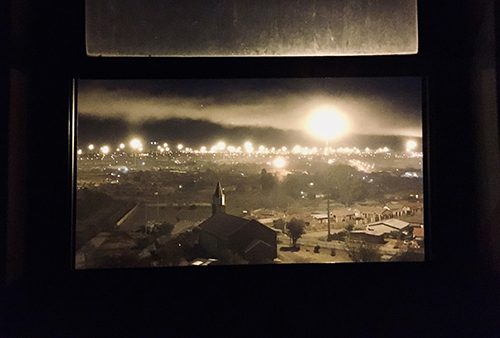
Janse van Rensburg’s late-night photo taken from the Pelonomi Hospital in Heidedal, Bloemfontein, shows the economic inequality that persists in South Africa. The photo was highly commended at the Imperial College London’s Global Creative Competition for Medical Student Responses to COVID-19.
A little shocked
He was a little shocked when he heard his photograph was highly commended. Janse van Rensburg says: “Imperial College London is a big institution and being an international competition I did not really expect a lot. There were participants from over 52 countries, and having seen some of the works that were submitted it feels special to be one of the students being noticed.”
Janse van Rensburg, who has never considered doing art, heard about the competition through the Faculty of Health Sciences platforms during lockdown level 5. He saw it as an opportunity to reflect, which has become even more imperative in times like these.
He says he does not go searching for art, but “notices” it from being conscious – something he thinks is important in medicine and life.
Value of creativity in promoting mental well-being
Dr Lynette van der Merwe, undergraduate medical programme director, School of Clinical Medicine, congratulated Janse van Rensburg, saying this commendation in an international competition underscores his talent and the value of creativity in promoting mental well-being.
“Heinrich’s artwork and showcase precisely what we aspire to develop in our exceptional UFS doctors-in-training: a professional with self-awareness, empathy and humanity.
“We initiated a Mental Health Awareness initiative and art competition in the School of Clinical Medicine in 2018 to promote creative expression as a means of supporting students’ mental health. Heinrich has won awards with his creative contributions every year, exhibiting his imaginative ability.”
Surgery and photography
Janse van Rensburg says he has always loved beautiful things and the meaning people attach to art is a good way to communicate that. He has applied for an internship at the Mitchells Plain hospital for when he completes his studies at the end of this year and is thinking of specialising in reconstructive or pediatrics surgery. Besides that, he would like to tap into his creative side and continue with the photography.
- Watch the video of the winners here