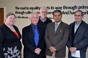
Attending the lecture were, from the left: Dr Chantel Swart, Prof Lodewyk Kock, Prof Debabrata Mukhopadhyay, Prof James du Preez; back: Prof Pieter van Wyk. Dr Swart, Profs Kock and Du Preez are from the Department of Microbial, Biochemical and Food Biotechnology. Prof Mukhopadhyay is from the Mayo Clinic (US) and Prof Van Wyk is from the Centre for Microscopy at the UFS.
Photo: Supplied |
The UFS made a discovery that may have enormous implications for the treatment of diseases in humans.
Since the discovery, the UFS joined forces with the Mayo Clinic in Rochester, US, in the fight against cancer.
In this collective effort, UFS researchers would be able to assist the Mayo team to:
• see how treatment in cancer patients is progressing,
• target treatments more effectively,
• reduce dosages in order to make treatment gentler on the patient,
• track the effectiveness of the chemotherapy drugs used, and
• gain an accurate view of how the cancer is being eliminated.
Prof Lodewyk Kock, Outstanding Professor at the Department of Microbial, Biochemical and Food Biotechnology, and his team incidentally created a technique to use argon gas particles for the first time on biological material to slice open cells to look inside.
The team that supported Prof Kock includes Dr Chantel Swart, Khumisho Dithebe (PhD student), Prof Hendrik Swart (Department of Physics) and Prof Pieter van Wyk (Centre for Microscopy).
Prof Debabrata Mukhopadhyay from the Mayo Clinic in Rochester, US, got to hear about this breakthrough at the UFS and a collaboration between the two institutions was established.
During a visit to the Bloemfontein Campus, Prof Mukhopadhyay explained novel techniques that make use of gold nanoparticles. These particles attach to chemotherapeutic drugs to selectively target cancer cells – dramatically decreasing the side effects to normal human cells.
For these new drugs (coupled to gold nanoparticles) to be accepted into clinical practice, visual and chemical proof is needed, though. This is where the technique developed by the UFS will play a vital role.
With the technique to look inside cells, the composition, location and metabolism of these drugs can be determined. This will aid in a proof of concept for the application of the nano-drugs. Furthermore, it will enable approval for use of these drugs in clinical trials and eventually could revolutionise cancer treatment as a whole.
For video lectures on the technique used, as well as its findings, follow these links:
1.
http://vimeo.com/63643628 (Comic version for school kids)
2.
http://vimeo.com/61521401 (Detailed version for fellow scientists)