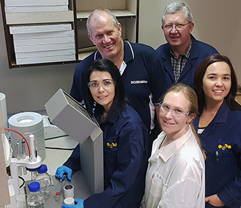
Inorganic Chemistry supervisors in the Radiopharmacy
Laboratory during the preparation of a typical complex
mixture to see how fast it reacts. Here are, from the left,
front: Dr Marietjie Schutte-Smith, Dr Alice Brink
(both scholars from the UFS Prestige
Scholar Programme), and Dr Truidie Venter (all three
are Thuthuka-funded researchers).
Back: Prof André Roodt and Dr Johan Venter.
Photo: Supplied
Imagine that you have been diagnosed with bone cancer and only have six months to live. You are in a wheelchair because the pain in your legs is so immense that you can’t walk anymore – similar to a mechanism eating your bones from the inside.
You are lucky though, since you could be injected with a drug to control the pain so effective that you will be able to get out of the wheelchair within a day-and-a-half and be able to walk again. Real-life incidents like these provide intense job satisfaction to Prof André Roodt, Head of Inorganic Chemistry at the University of the Free State (UFS). The research, which is conducted by the Inorganic Group at the UFS, contributes greatly to the availability of pain therapy that does not involve drugs, but improves the quality of life for cancer patients.
The research conducted by the Inorganic Group under the leadership of Prof Roodt, plays a major role in the clever design of model medicines to better detect and treat cancer.
The Department of Chemistry is one of approximately 10 institutions worldwide that conducts research on chemical mechanisms to identify and control cancer. “The fact that we are able to cooperate with the Departments of Nuclear Medicine and Medical Physics at the UFS, the Animal Research Centre, and other collaborators in South Africa and abroad, but especially the methodology we utilise to conduct research (studying the chemical manner in which drugs are absorbed in cancer as well as the time involved), enhances the possibility of making a contribution to cancer research,” says Prof Roodt.
Technique to detect cancer spots on bone
According to the professor, there are various ways of detecting cancer in the body. Cancer can, inter alia, be identified by analysing blood, X-rays (external) or through an internal technique where the patient is injected with a radioactive isotope.
Prof Roodt explains: “The doctor suspects that the patient has bone cancer and injects the person with a drug consisting of an isotope (only emits X-rays and does no damage to tissue) that is connected to a phosphonate (similar to those used for osteoporosis). Once the drug is injected, the isotope (Technetium-99m) moves to the spot on the bone where the cancer is located. The gamma rays in the isotope illuminate the area and the doctor can see exactly where treatment should be applied. The Technetium-99m has the same intensity gamma rays as normal X-rays and therefore operates the same as an internal X-ray supply.” With this technique, the doctor can see where the cancer spots are within a few hours.
The same technique can be used to identify inactive parts of the brain in Alzheimer patients, as well as areas of the heart where there is no blood supply or where the heart muscle is dead.
Therapeutic irradiation of cancer
For the treatment of pain connected with cancer, the isotope Rhenium-186 is injected. Similar to the manner in which the Technetium-99m phosphonate compound is ingested into the body, the Rhenium-186 phosphonate travels to the cancer spots. Patients thus receive therapeutic irradiation – a technique known as palliative therapy, which is excellent for treating pain. A dosage of this therapy usually lasts for about two months.
The therapy is, however, patient specific. The dosages should correspond with the occurrence and size of cancer spots in the patient’s body. First, the location of the cancer will be determined by means of a technetium scan. After that, the size of the area where the cancer occurs has to be determined. The dosage for addressing total pain distribution will be calculated according to these results.
Technique to detect cancer spots on soft tissue
Another technique to detect cancer as spots on bone or in soft tissue and organs throughout the body is by utilising a different type of irradiation, a so-called PET isotope. The Fluor-18 isotope is currently used widely, and in Pretoria a machine called a cyclotron was produced by Dr Gerdus Kemp, who is a former PhD graduate from the Inorganic Research Group. The F-18 is then hidden within a glucose molecule and a patient will be injected with the drug after being tranquillised and after the metabolism has been lowered considerably. The glucose, which is the ‘food' that cancer needs to grow, will then travel directly to the cancer area and the specific area where the cancer is located will thus be traced and ‘illuminated’ by the Fluor-18, which emits its own 'X-rays'.
In the late 80s, Prof Roodt did his own postdoctoral study on this research in the US. He started collaborating with the Department of Nuclear Medicine at the UFS in the early 90s, when he initiated testing for this research.
Through their research of more than 15 years, the Inorganic Group in the Department of Chemistry has made a major contribution to cancer research. Research on mechanisms for the detection of cancer, by designing new clever chemical agents, and the chemical ways in which these agents are taken up in the body, especially contributes to the development in terms of cancer therapy and imaging, and has been used by a number of hospitals in South Africa.
The future holds great promise
Prof Roodt and his team are already working on a bilateral study between the UFS and Kenya. It involves the linking of radio isotopes, as mentioned above, to known natural products (such as rooibos tea), which possess anti-cancer qualities.