Latest News Archive

Please select Category, Year, and then Month to display items
Previous Archive
27 July 2021 | Story Leonie Bolleurs | Photo UFS Photo Archive
Prof Hendrik Swart played a key role in the Department of Physics acquiring the PHI Quantes XPS system, the first in Africa and one of only 20 in the world.

The state-of-the-art equipment in the Department of Physics at the University of the Free State (UFS) differentiates this department from its competitors. Availability of the equipment makes it possible for researchers as well as students to deliver work that receives national and international recognition. 

Recently, the department acquired a PHI Quantes XPS system, the first in Africa and one of only 20 in the world. 

Creating better phosphor 

“The Quantes XPS system uses X-rays to determine the chemical composition of molecules on the surface of a sample. The system is unique in the sense that it also has an extra X-ray source that can determine the chemical state below the surface, which was not possible in the past.  This will help us to dictate the position of defects in our phosphor materials that will consequently enable us to create better phosphor for solid state lighting as well as solar cell applications,” explains Prof Hendrik Swart, Senior Professor in the Department of Physics, who also holds the SARChI Chair in Solid State Luminescent and Advanced Materials.

After he had the opportunity to observe the system in the factory in Chigasaki, Kanagawa, Japan, where he attended a conference, Prof Swart was very impressed by its performance. He discussed it with Prof Koos Terblans, Head of the department, and other colleagues, and started making plans to buy the system. 

When the department first bought the X-ray photoelectron spectroscopy (XPS) system in 2007/2008, it became the national facility on XPS measurements. Not only is this an upgrade of the XPS system bought 14 years ago, but the new system will enable the department to do more measurements. “The number of samples that we have to handle has just become too much for one system. The new system’s increased capacity for making measurements addresses this challenge and it also gives UFS scientists and postgraduate students more time to spend on fundamental measurements to develop research of a higher level,” says Prof Swart.

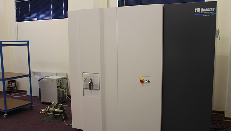
(The Quantes XPS system. Photo:Supplied)

Explaining about the measurements, Prof Swart says: “This advanced X-ray photoelectron spectroscopy (XPS) instrument has the capability to analyse the very small area that the user is interested in and a large area of the uniform sample surface. The two different types of X-ray sources – the hard X-ray source and the more conventional soft X-ray source – can be switched automatically, allowing users to analyse the same area and/or points of a sample. The PHI Quantes XPS system ensures the availability of superior features such as automatic analysis, automatic sample transfer, turnkey charge neutralisation, and advanced data processing.”

“This XPS instrument is designed to pioneer new methods and applications transcending conventional ideas of what is possible.”

Optimising efficiency of materials

Prof Swart says the Department of Physics, especially the Research Chair in Advanced and Luminescent Materials, is developing new high-technology materials on a daily basis. “It is very important to know the chemical composition and defect distribution of the materials in order to add value to the fabrication of these materials,” he adds.

“The distribution of these defects is vital for the efficiency of the phosphor materials. If we know where these defects are located, we can determine the mechanisms of the light output coming from these phosphors,” describes Prof Swart.

Research conducted as part of the Research Chair in Solid State Luminescent and Advanced Materials will benefit significantly from this new system.

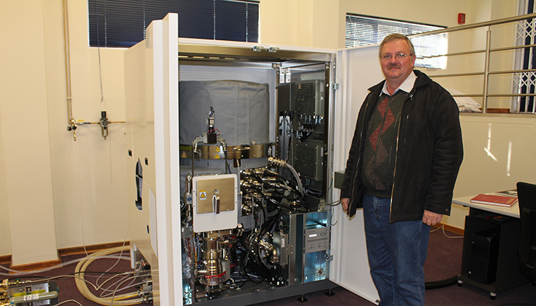
(Prof Koos Terblans, Head of the Department of Physics next to the Quantes XPS system. Photo:Supplied) 

“We are currently concentrating on phosphors as sensors (temperature), light-emitting diodes (LEDs), and solar cells, where we optimise the efficiency and durability of these materials. Any new knowledge, which I believe the PHI Quantes XPS system will provide us, will help us to reach our goal much quicker,” he says. 

Apart from the positive impact on research, the PHI Quantes XPS system will also be a benefit to society in the long term. Improved LEDs can be used to save electricity, and better solar cells can help to generate electricity, to mention but two examples. 

News Archive

Nanotechnology breakthrough at UFS
2010-08-19

 Ph.D students, Chantel Swart and Ntsoaki Leeuw


Scientists at the University of the Free State (UFS) made an important breakthrough in the use of nanotechnology in medical and biological research. The UFS team’s research has been accepted for publication by the internationally accredited Canadian Journal of Microbiology.

The UFS study dissected yeast cells exposed to over-used cooking oil by peeling microscopically thin layers off the yeast cells through the use of nanotechnology.

The yeast cells were enlarged thousands of times to study what was going on inside the cells, whilst at the same time establishing the chemical elements the cells are composed of. This was done by making microscopically small surgical incisions into the cell walls.

This groundbreaking research opens up a host of new uses for nanotechnology, as it was the first study ever in which biological cells were surgically manipulated and at the same time elemental analysis performed through nanotechnology. According to Prof. Lodewyk Kock, head of the Division Lipid Biotechnology at the UFS, the study has far reaching implications for biological and medical research.

The research was the result of collaboration between the Department of Microbial, Biochemical and Food Biotechnology, the Department of Physics (under the leadership of Prof. Hendrik Swart) and the Centre for Microscopy (under the leadership of Prof.Pieter van Wyk).

Two Ph.D. students, Chantel Swart and Ntsoaki Leeuw, overseen by professors Kock and Van Wyk, managed to successfully prepare yeast that was exposed to over-used cooking oil (used for deep frying of food) for this first ever method of nanotechnological research.

According to Prof. Kock, a single yeast cell is approximately 5 micrometres long. “A micrometre is one millionth of a metre – in laymen’s terms, even less than the diameter of a single hair – and completely invisible to the human eye.”

Through the use of nanotechnology, the chemical composition of the surface of the yeast cells could be established by making a surgical incision into the surface. The cells could be peeled off in layers of approximately three (3) nanometres at a time to establish the effect of the oil on the yeast cell’s composition. A nanometre is one thousandth of a micrometre.

Each cell was enlarged by between 40 000 and 50 000 times. This was done by using the Department of Physics’ PHI700 Scanning Auger Nanoprobe linked to a Scanning Electron Microscope and Argon-etching. Under the guidance of Prof. Swart, Mss. Swart en Leeuw could dissect the surfaces of yeast cells exposed to over-used cooking oil. 

The study noted wart like outgrowths - some only a few nanometres in diameter – on the cell surfaces. Research concluded that these outgrowths were caused by the oil. The exposure to the oil also drastically hampered the growth of the yeast cells. (See figure 1)  

Researchers worldwide have warned about the over-usage of cooking oil for deep frying of food, as it can be linked to the cause of diseases like cancer. The over-usage of cooking oil in the preparation of food is therefore strictly regulated by laws worldwide.

The UFS-research doesn’t only show that over-used cooking oil is harmful to micro-organisms like yeast, but also suggests how nanotechnology can be used in biological and medical research on, amongst others, cancer cells.


Figure 1. Yeast cells exposed to over-used cooking oil. Wart like protuberances/ outgrowths (WP) is clearly visible on the surfaces of the elongated yeast cells. With the use of nanotechnology, it is possible to peel off the warts – some with a diameter of only a few nanometres – in layers only a few nanometres thick. At the same time, the 3D-structure of the warts as well as its chemical composition can be established.  

Media Release
Issued by: Mangaliso Radebe
Assistant Director: Media Liaison
Tel: 051 401 2828
Cell: 078 460 3320
E-mail: radebemt@ufs.ac.za  
18 August 2010
 

We use cookies to make interactions with our websites and services easy and meaningful. To better understand how they are used, read more about the UFS cookie policy. By continuing to use this site you are giving us your consent to do this.

Accept