Latest News Archive

Please select Category, Year, and then Month to display items
Previous Archive
25 April 2022 | Story Elsabé Brits
Andre Roodt and Alice Brink
Prof Andreas Roodt and Prof Alice Brink are two of the inventors of the ‘Multinuclear complexes and their preparation patent.

According to the World Health Organisation (WHO), cancer is a leading cause of death worldwide, accounting for nearly 10 million deaths in 2020, or nearly one in six. The most common cancers are breast, lung, colon, rectum, and prostate cancers. There is a constant need to provide methods to diagnose and treat cancer-related tumours.  Current research strategies focus on eliminating cancer cells with the minimum damage to surrounding healthy cells.

A limitation of current technologies is that they are mostly based on the separate identification of cancer (diagnostic), followed by treatment (therapy) using chemotherapy and/or radiotherapy. To fit both needs at the same time and with similar or identical compounds, the principle of theranostic medicine was identified. This concept employs both diagnosing (by imaging) cancer and delivering therapy (treatment) simultaneously, which has been receiving increased attention internationally.

Collaborating with the University of Zurich
A University of the Free State (UFS) team, together with a team from the University of Zürich, conducted exciting research in this area and filed a patent titled ‘Multinuclear complexes and their preparation’. The patent was granted in South Africa and by the European Patent Office. It is being validated in selected European countries. The patent is pending in the USA, Japan, Hong Kong, and India. The inventors from the UFS are Prof Andreas Roodt, Prof Alice Brink, Dr Pennie Mokolokolo, and Dr Vincent Dumisani Kama. The approach that their technology takes is to enable the synthesis of a multinuclear compound/s, which may contain different pre-selected radioisotopes, to allow both imaging and therapy to the cancer site(s) with one and the same metal-organic complex.

So far, high-yield production of compounds has been successfully innovated, which contain both an imaging (in particular the widely utilised imaging isotope Technetium-99m) and therapeutic (typically the therapeutic isotope Rhenium-186) radioactive isotope(s), optionally carrying an additional cytotoxic agent. (Chemotherapy uses anti-cancer [cytotoxic] drugs to destroy cancer cells.)

Nuclear medicine technologies
In the next phase of the research, a lead compound portfolio of four to five model pharmaceuticals containing these metal nuclides with appropriate directing groups to target cancer sites will be designed and constructed. A number of these entities are known and can be introduced through different techniques. These will then undergo full characterisation and efficacy evaluation in biological models (in vitro), followed by extensive animal and human trials.

The technology will be delivered as a product or service in the way that current nuclear medicine technologies are delivered.

The fact that this product(s) contains both imaging and therapeutic radionuclides or cytotoxic modalities, enables detailed tracking of the pharmaceutical and monitoring of the tumours' response to the therapy. Not directly related to the patent, but an asset to it, is the fact that the incorporation of rhenium with a high atomic number (Z = 75) opens the additional opportunity to utilise the multinuclear compounds also as radiosensitisers. Synergistic effects, enhancing the therapeutic efficacy, can thus be expected in combination with radiotherapy.

The UFS would like to partner with a pharmaceutical company working in the field of nuclear medicine to commercialise this technology. Interested parties can contact Ravini Moodley at MoodleyR5@ufs.ac.za

News Archive

Nanotechnology breakthrough at UFS
2010-08-19

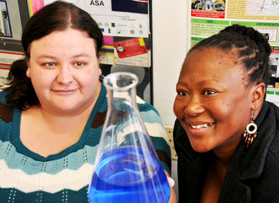
 Ph.D students, Chantel Swart and Ntsoaki Leeuw


Scientists at the University of the Free State (UFS) made an important breakthrough in the use of nanotechnology in medical and biological research. The UFS team’s research has been accepted for publication by the internationally accredited Canadian Journal of Microbiology.

The UFS study dissected yeast cells exposed to over-used cooking oil by peeling microscopically thin layers off the yeast cells through the use of nanotechnology.

The yeast cells were enlarged thousands of times to study what was going on inside the cells, whilst at the same time establishing the chemical elements the cells are composed of. This was done by making microscopically small surgical incisions into the cell walls.

This groundbreaking research opens up a host of new uses for nanotechnology, as it was the first study ever in which biological cells were surgically manipulated and at the same time elemental analysis performed through nanotechnology. According to Prof. Lodewyk Kock, head of the Division Lipid Biotechnology at the UFS, the study has far reaching implications for biological and medical research.

The research was the result of collaboration between the Department of Microbial, Biochemical and Food Biotechnology, the Department of Physics (under the leadership of Prof. Hendrik Swart) and the Centre for Microscopy (under the leadership of Prof.Pieter van Wyk).

Two Ph.D. students, Chantel Swart and Ntsoaki Leeuw, overseen by professors Kock and Van Wyk, managed to successfully prepare yeast that was exposed to over-used cooking oil (used for deep frying of food) for this first ever method of nanotechnological research.

According to Prof. Kock, a single yeast cell is approximately 5 micrometres long. “A micrometre is one millionth of a metre – in laymen’s terms, even less than the diameter of a single hair – and completely invisible to the human eye.”

Through the use of nanotechnology, the chemical composition of the surface of the yeast cells could be established by making a surgical incision into the surface. The cells could be peeled off in layers of approximately three (3) nanometres at a time to establish the effect of the oil on the yeast cell’s composition. A nanometre is one thousandth of a micrometre.

Each cell was enlarged by between 40 000 and 50 000 times. This was done by using the Department of Physics’ PHI700 Scanning Auger Nanoprobe linked to a Scanning Electron Microscope and Argon-etching. Under the guidance of Prof. Swart, Mss. Swart en Leeuw could dissect the surfaces of yeast cells exposed to over-used cooking oil. 

The study noted wart like outgrowths - some only a few nanometres in diameter – on the cell surfaces. Research concluded that these outgrowths were caused by the oil. The exposure to the oil also drastically hampered the growth of the yeast cells. (See figure 1)  

Researchers worldwide have warned about the over-usage of cooking oil for deep frying of food, as it can be linked to the cause of diseases like cancer. The over-usage of cooking oil in the preparation of food is therefore strictly regulated by laws worldwide.

The UFS-research doesn’t only show that over-used cooking oil is harmful to micro-organisms like yeast, but also suggests how nanotechnology can be used in biological and medical research on, amongst others, cancer cells.
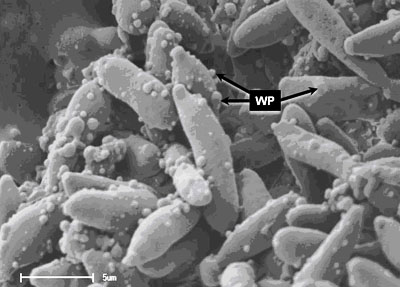
 

Figure 1. Yeast cells exposed to over-used cooking oil. Wart like protuberances/ outgrowths (WP) is clearly visible on the surfaces of the elongated yeast cells. With the use of nanotechnology, it is possible to peel off the warts – some with a diameter of only a few nanometres – in layers only a few nanometres thick. At the same time, the 3D-structure of the warts as well as its chemical composition can be established.  

Media Release
Issued by: Mangaliso Radebe
Assistant Director: Media Liaison
Tel: 051 401 2828
Cell: 078 460 3320
E-mail: radebemt@ufs.ac.za  
18 August 2010
 

We use cookies to make interactions with our websites and services easy and meaningful. To better understand how they are used, read more about the UFS cookie policy. By continuing to use this site you are giving us your consent to do this.

Accept