Latest News Archive
Please select Category, Year, and then Month to display items
01 March 2023
|
Story Alicia Pienaar
 Prof Mathys Labuschagne is the Head of the Clinical Simulation and Skills Unit within the School of Biomedical Sciences in the Faculty of Health Sciences
The Dean of the Faculty of Health Sciences, Prof Gert van Zyl, invites you to the inaugural lecture of Prof Mathys Labuschagne, Head of the Clinical Simulation and Skills Unit within the School of Biomedical Sciences in the Faculty of Health Sciences.
Subject: Clinical Simulation: Quo Vadis?
Venue: Auditorium, Equitas Building, Bloemfontein Campus
Date: 8 March 2023
Time: 17:30
RSVP on or before Friday 3 March 2023
Light refreshments will be served after the inaugural lecture.
About Prof M Labuschagne
Prof Mathys Labuschagne is the Head of the Clinical Simulation and Skills Unit within the School of Biomedical Sciences in the Faculty of Health Sciences at the University of the Free State. He completed his MB ChB degree and qualified as an ophthalmologist in 2006.
He developed an interest in health professions education and obtained a PhD in Health Professions Education in June 2012. The title of his thesis was: Clinical Simulation to enhance undergraduate medical education and training at the University of the Free State.
Prof Labuschagne was appointed Head of the Clinical Simulation and Skills Unit at the University of the Free State. The facility is utilised for undergraduate and postgraduate clinical simulation training, as well as interprofessional training and research. He has a special interest in simulation as training tool, precision skills training, and mastery of learning and simulation as tool to prepare students for interprofessional education and collaborative practice.
Prof Labuschagne is part of a multi-institutional consortium that developed the MySkills Medic app. It is a clinical procedural skills application aimed at graduating medical students, interns, and community-service doctors. He was appointed as a member of the Ophthalmology Foundation Education Simulation Subcommittee (affiliate of the International Council of Ophthalmology) tasked with developing a white paper to guide simulation training for ophthalmologists. He is involved in postgraduate supervision for master’s and PhD students in HPE.
Mathematical methods used to detect and classify breast cancer masses
2016-08-10
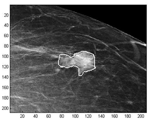 Examples of Acho’s breast mass
segmentation identification
Breast cancer is the leading cause of female mortality in developing countries. According to the World Health Organization (WHO), the low survival rates in developing countries are mainly due to the lack of early detection and adequate diagnosis programs.
Seeing the picture more clearly
Susan Acho from the University of the Free State’s Department of Medical Physics, breast cancer research focuses on using mathematical methods to delineate and classify breast masses. Advancements in medical research have led to remarkable progress in breast cancer detection, however, according to Acho, the methods of diagnosis currently available commercially, lack a detailed finesse in accurately identifying the boundaries of breast mass lesions.
Inspiration drawn from pioneer
Drawing inspiration from the Mammography Computer Aided Diagnosis Development and Implementation (CAADI) project, which was the brainchild Prof William Rae, Head of the department of Medical Physics, Acho’s MMedSc thesis titled ‘Segmentation and Quantitative Characterisation of Breast Masses Imaged using Digital Mammography’ investigates classical segmentation algorithms, texture features and classification of breast masses in mammography. It is a rare research topic in South Africa.
Characterisation of breast masses, involves delineating and analysing the breast mass region on a mammogram in order to determine its shape, margin and texture composition. Computer-aided diagnosis (CAD) program detects the outline of the mass lesion, and uses this information together with its texture features to determine the clinical traits of the mass. CAD programs mark suspicious areas for second look or areas on a mammogram that the radiologist might have overlooked. It can act as an independent double reader of a mammogram in institutions where there is a shortage of trained mammogram readers.
Light at the end of the tunnel
Breast cancer is one of the most common malignancies among females in South Africa. “The challenge is being able to apply these mathematical methods in the medical field to help find solutions to specific medical problems, and that’s what I hope my research will do,” she says.
By using mathematics, physics and digital imaging to understand breast masses on mammograms, her research bridges the gap between these fields to provide algorithms which are applicable in medical image interpretation.