Latest News Archive

Please select Category, Year, and then Month to display items
Previous Archive
23 March 2023 | Story Rulanzen Martin | Photo Stephen Collett
From left: Hanlie Grobler, Senior Officer at the CFM; Prof Koos Terblans, Head of the Physics Department; Nonkululeko Phili, Assistant Officer at the CFM; and Edward Lee, Junior Lecturer and Researcher at the CFM. Photo: Stephen Collett

The 
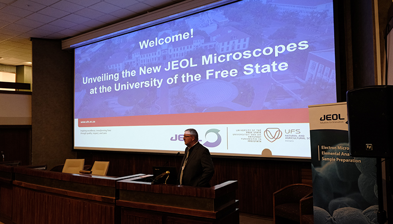
Centre for Microscopy (CFM) in the Faculty of Natural and Agricultural Sciences at the University of the Free State (UFS) unveiled a sophisticated JEOL High Resolution Transmission Electron Microscope (HRTEM) during a two-day microscopy conference on 14 and 15 March 2023. The microscope is part of a larger investment into research equipment worth R65 million. 

Speaking at the opening of the conference, Prof Corli Witthuhn, out-going Vice-Rector: Research and Internationalisation, said the microscope purchase “is a significant milestone in the university’s bid for cutting-edge research”. The HRTEM is part of a larger consignment of JEOL equipment at the UFS and, according to Dr Sarah Harper from JEOL UK, it places the UFS in a unique position.  

UFS at the forefront in using electron microscopes  

The HRTEM microscope can be utilised across disciplines and will give the UFS an advantage in uncovering new solutions and creating national and international interdisciplinary research collaborations. “The UFS is at the forefront in this field in SA and continues to push the boundaries,” Prof Witthuhn said. This move will also positively impact the training of honours, master’s, and doctoral students. 

Prof Danie Vermeulen, Dean of the Faculty of Natural and Agricultural Sciences, reiterated Prof Witthuhn’s sentiments by saying that this equipment will set the faculty apart from its competitors. “The faculty already reached the goals of Vision 130 by being proactive,” he said. In the past seven years more than R300 million worth of equipment was acquired by the faculty, but he added that to be the best is not just about the best equipment – “the data coming from using this equipment is what will make the real difference”.

Prof Koos Terblans
Prof Koos Terblans opens the conference on 14 March 2023. Photo: Stephen Collett .

Road to the JEOL HRTEM started in 2018

The process of acquiring a HRTEM microscope started in 2018 and was concluded with the purchasing of the JOEL microscopes in March 2020, a few weeks before the first COVID-19 lockdown. The purchase was made possible through the collaboration between the faculties of Natural and Agricultural Sciences and Health Sciences. Thanks to the dedication of staff members in the Centre for Microscopy and Physics, it was possible to accept delivery of the new HRTEM in June 2021. Prof Koos Terblans, Head of the Physics Department and the Centre for Microscopy, who led the entire project, said this was one of the “proudest moments in my career”.  

Installing the equipment involved various university resources, including the University Estates Department, which had to make additional structural changes to the room where the equipment is housed. This included digging two metres into the existing floor and placing the HRTEM on a 70-tonne solid concrete block, to ensure that the equipment was secure and vibration free.

Prof Terblans said now that the HRTEM from JEOL and its supporting equipment – the final piece of the R65 million research investment puzzle – is part of the faculty’s resources, it is up to the scientists and academics to utilise it for innovative research, enhance research productivity, and foster new collaborations. 

Edward Lee
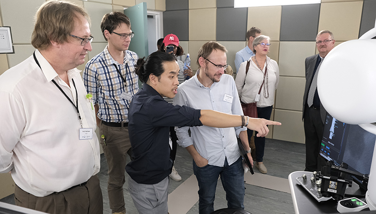
Edward Lee shows the new HRTEM electron microscope to colleagues and conference attendees.Photo: Stephen Collett 

News Archive

Art and science help us understand the world and our place in it
2017-10-28


Description: Art and science  Tags: Art and science

At the event were, from the left: Tristan Nel, first-year Fine Arts student;
Dr Janine Allen-Spies from the Department of Fine Arts;
Prof Carlien Pohl-Albertyn from the Department of Microbiology,
Biochemical and Food Biotechnology; and Pheny Mokawane, a
Microbiology, Biochemical and Food Biotechnology student.
Photo: Charl Devenish

Although BioArt dates back as far as the 15th and 16th centuries with the work of Leonardo da Vinci, it is not every day that art and science combine. This rare phenomenon made its appearance when two totally different groups of students – studying arts and microbiology respectively – joined hands in an initiative to create BioArt.

This first-time undergraduate teaching collaboration between the Departments of Fine Arts and Microbial, Biochemical and Food Biotechnology at the University of the Free State (UFS), which is characterised by the use of living materials, such as enzymes, microbes and DNA, as well as scientific tools and methods, is exploring a number of questions. 

Different outcomes for arts and microbiology students

According to Prof Carlien Pohl-Albertyn from the Department of Microbiology, Biochemical and Food Biotechnology, one of the central questions explored in BioArt is the nature of ‘life’. “At which stage can matter be classified as being alive or living?” she asked. 

“We realised that the outcomes for the two groups of students would not be the same. For the microbiology students, the focus would be on the understanding and effective communication of a microbiological concept. For the art students the focus would be on the execution of the assignment using visual elements and applied theory of art,” said Prof Pohl-Albertyn.

Dr Janine Allen-Spies from the Department of Fine Arts added: “Art students will also be exploring strangely or previously unforeseen gaps between art and science that can be filled with imaginative interpretations which may forward creative insights in both BioArt as a developing art form and microbiology as investigative science.”

Students’ understanding of microbial evolution reflected in art
The art students had to visit the microbiology labs for their assignment as this is mostly a foreign environment for these students. “The paint medium they had to use was gouache. This medium with its bright colours works well to depict microscopic organisms in art,” Dr Allen-Spies said. 

On display at the Department of Microbial, Biochemical and Food Biotechnology on the Bloemfontein Campus, at a recent event to introduce this new initiative to a wider audience, was a range of visually and scientifically compelling paintings and artefacts (such as paintings, poems, songs, apps) which explore a theme within microbiology from a BioArt perspective that uses creativity to communicate concepts dealt with in the module Microbial Evolution and Diversity.

Any parties who are interested in buying the art can contact Dr Allen-Spies at allenj@ufs.ac.za.

Paintings and artefacts reflects students understanding of BioArt. At the recent opening of the BioArt exhibition at the UFS Department of Microbial, Biochemical and Food Biotechnology, was the work of Madeleen Jansen van Rensburg on display.

Pheny Mokawane, a Microbiology, Biochemical and Food Biotechnology student, wrote a poem for his BioArt project in the Microbial Evolution and Diversity assignment. 


We use cookies to make interactions with our websites and services easy and meaningful. To better understand how they are used, read more about the UFS cookie policy. By continuing to use this site you are giving us your consent to do this.

Accept