Latest News Archive

Please select Category, Year, and then Month to display items
Previous Archive
23 March 2023 | Story Rulanzen Martin | Photo Stephen Collett
From left: Hanlie Grobler, Senior Officer at the CFM; Prof Koos Terblans, Head of the Physics Department; Nonkululeko Phili, Assistant Officer at the CFM; and Edward Lee, Junior Lecturer and Researcher at the CFM. Photo: Stephen Collett

The Centre for Microscopy (CFM) in the Faculty of Natural and Agricultural Sciences at the University of the Free State (UFS) unveiled a sophisticated JEOL High Resolution Transmission Electron Microscope (HRTEM) during a two-day microscopy conference on 14 and 15 March 2023. The microscope is part of a larger investment into research equipment worth R65 million. 

Speaking at the opening of the conference, Prof Corli Witthuhn, out-going Vice-Rector: Research and Internationalisation, said the microscope purchase “is a significant milestone in the university’s bid for cutting-edge research”. The HRTEM is part of a larger consignment of JEOL equipment at the UFS and, according to Dr Sarah Harper from JEOL UK, it places the UFS in a unique position.  

UFS at the forefront in using electron microscopes  

The HRTEM microscope can be utilised across disciplines and will give the UFS an advantage in uncovering new solutions and creating national and international interdisciplinary research collaborations. “The UFS is at the forefront in this field in SA and continues to push the boundaries,” Prof Witthuhn said. This move will also positively impact the training of honours, master’s, and doctoral students. 

Prof Danie Vermeulen, Dean of the Faculty of Natural and Agricultural Sciences, reiterated Prof Witthuhn’s sentiments by saying that this equipment will set the faculty apart from its competitors. “The faculty already reached the goals of Vision 130 by being proactive,” he said. In the past seven years more than R300 million worth of equipment was acquired by the faculty, but he added that to be the best is not just about the best equipment – “the data coming from using this equipment is what will make the real difference”.

Prof Koos Terblans
Prof Koos Terblans opens the conference on 14 March 2023. Photo: Stephen Collett .

Road to the JEOL HRTEM started in 2018

The process of acquiring a HRTEM microscope started in 2018 and was concluded with the purchasing of the JOEL microscopes in March 2020, a few weeks before the first COVID-19 lockdown. The purchase was made possible through the collaboration between the faculties of Natural and Agricultural Sciences and Health Sciences. Thanks to the dedication of staff members in the Centre for Microscopy and Physics, it was possible to accept delivery of the new HRTEM in June 2021. Prof Koos Terblans, Head of the Physics Department and the Centre for Microscopy, who led the entire project, said this was one of the “proudest moments in my career”.  

Installing the equipment involved various university resources, including the University Estates Department, which had to make additional structural changes to the room where the equipment is housed. This included digging two metres into the existing floor and placing the HRTEM on a 70-tonne solid concrete block, to ensure that the equipment was secure and vibration free.

Prof Terblans said now that the HRTEM from JEOL and its supporting equipment – the final piece of the R65 million research investment puzzle – is part of the faculty’s resources, it is up to the scientists and academics to utilise it for innovative research, enhance research productivity, and foster new collaborations. 

Edward Lee
Edward Lee shows the new HRTEM electron microscope to colleagues and conference attendees.Photo: Stephen Collett 

News Archive

Nanotechnology breakthrough at UFS
2010-08-19

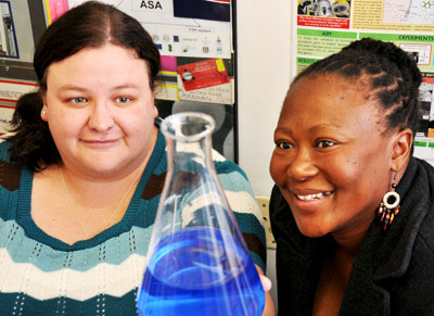
 Ph.D students, Chantel Swart and Ntsoaki Leeuw


Scientists at the University of the Free State (UFS) made an important breakthrough in the use of nanotechnology in medical and biological research. The UFS team’s research has been accepted for publication by the internationally accredited Canadian Journal of Microbiology.

The UFS study dissected yeast cells exposed to over-used cooking oil by peeling microscopically thin layers off the yeast cells through the use of nanotechnology.

The yeast cells were enlarged thousands of times to study what was going on inside the cells, whilst at the same time establishing the chemical elements the cells are composed of. This was done by making microscopically small surgical incisions into the cell walls.

This groundbreaking research opens up a host of new uses for nanotechnology, as it was the first study ever in which biological cells were surgically manipulated and at the same time elemental analysis performed through nanotechnology. According to Prof. Lodewyk Kock, head of the Division Lipid Biotechnology at the UFS, the study has far reaching implications for biological and medical research.

The research was the result of collaboration between the Department of Microbial, Biochemical and Food Biotechnology, the Department of Physics (under the leadership of Prof. Hendrik Swart) and the Centre for Microscopy (under the leadership of Prof.Pieter van Wyk).

Two Ph.D. students, Chantel Swart and Ntsoaki Leeuw, overseen by professors Kock and Van Wyk, managed to successfully prepare yeast that was exposed to over-used cooking oil (used for deep frying of food) for this first ever method of nanotechnological research.

According to Prof. Kock, a single yeast cell is approximately 5 micrometres long. “A micrometre is one millionth of a metre – in laymen’s terms, even less than the diameter of a single hair – and completely invisible to the human eye.”

Through the use of nanotechnology, the chemical composition of the surface of the yeast cells could be established by making a surgical incision into the surface. The cells could be peeled off in layers of approximately three (3) nanometres at a time to establish the effect of the oil on the yeast cell’s composition. A nanometre is one thousandth of a micrometre.

Each cell was enlarged by between 40 000 and 50 000 times. This was done by using the Department of Physics’ PHI700 Scanning Auger Nanoprobe linked to a Scanning Electron Microscope and Argon-etching. Under the guidance of Prof. Swart, Mss. Swart en Leeuw could dissect the surfaces of yeast cells exposed to over-used cooking oil. 

The study noted wart like outgrowths - some only a few nanometres in diameter – on the cell surfaces. Research concluded that these outgrowths were caused by the oil. The exposure to the oil also drastically hampered the growth of the yeast cells. (See figure 1)  

Researchers worldwide have warned about the over-usage of cooking oil for deep frying of food, as it can be linked to the cause of diseases like cancer. The over-usage of cooking oil in the preparation of food is therefore strictly regulated by laws worldwide.

The UFS-research doesn’t only show that over-used cooking oil is harmful to micro-organisms like yeast, but also suggests how nanotechnology can be used in biological and medical research on, amongst others, cancer cells.
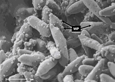
 

Figure 1. Yeast cells exposed to over-used cooking oil. Wart like protuberances/ outgrowths (WP) is clearly visible on the surfaces of the elongated yeast cells. With the use of nanotechnology, it is possible to peel off the warts – some with a diameter of only a few nanometres – in layers only a few nanometres thick. At the same time, the 3D-structure of the warts as well as its chemical composition can be established.  

Media Release
Issued by: Mangaliso Radebe
Assistant Director: Media Liaison
Tel: 051 401 2828
Cell: 078 460 3320
E-mail: radebemt@ufs.ac.za  
18 August 2010
 

We use cookies to make interactions with our websites and services easy and meaningful. To better understand how they are used, read more about the UFS cookie policy. By continuing to use this site you are giving us your consent to do this.

Accept