Latest News Archive
Please select Category, Year, and then Month to display items
29 March 2023
|
Story Samkelo Fetile
|
Photo Simba Matema
 From left to right: Prof Jean Bernard Lekana-Douki, Director General at the CIRMF; Prof Francis Petersen, Rector and Vice-Chancellor of the UFS; and Prof Jean-Fabrice Yala, Scientific and Technical Director at the CIRMF.
The University of the Free State (UFS) recently welcomed senior members from the International Centre for Medical Research in Franceville (CIRMF), Gabon to sign a Memorandum of Understanding (MoU) between the two entities.
CIRMF is a non-profit medical research centre that was established in 1974. Its primary focus is on diagnosing infectious diseases that pose immediate problems in Gabon and the Central African sub-region. The centre is dedicated to improving public health through various initiatives, including the training of Gabonese health executives in doctoral and post-doctoral education.
“The signing of the MoU with CIRMF is a step in the right direction as the UFS continues to strengthen its footprint on the African Continent. This is seminal in driving the university’s internationalisation processes and opening an avenue to expanding research networks, especially in the African continent,” said Bonolo Makhalemele, Coordinator of strategic projects at the Office for International Affairs, UFS.
In the interest of developing broader research collaborations, the CIRMF delegation met with some UFS academics in a series of engagements, particularly in the Health Sciences Faculty and the Natural and Agricultural Sciences Faculty. “The integration of knowledge and practices that emanate from the continent provides opportunities for the co-creation of knowledge that can be shared with the rest of the world,” said Kagiso Ngake, Coordinator for Partnerships, Collaborative Degrees, and Outgoing Mobility at the Office for International Affairs, UFS.
A concrete roadmap was established to formalise the collaborations that resulted from these engagements and will commence as soon as April 2023. “We have laid a solid foundation and are on the way to a strong and rich partnership!” exclaimed Prof Jean Bernard Lekana-Douki, Director General at the CIRMF.
Prof Francis Petersen, Rector, and Vice-Chancellor of the UFS, further highlighted how the newly established partnership with the CIRMF aligns with the mandate of the UFS Vision 130 of expanding the UFS Africa research network.
Mathematical methods used to detect and classify breast cancer masses
2016-08-10
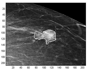 Examples of Acho’s breast mass
segmentation identification
Breast cancer is the leading cause of female mortality in developing countries. According to the World Health Organization (WHO), the low survival rates in developing countries are mainly due to the lack of early detection and adequate diagnosis programs.
Seeing the picture more clearly
Susan Acho from the University of the Free State’s Department of Medical Physics, breast cancer research focuses on using mathematical methods to delineate and classify breast masses. Advancements in medical research have led to remarkable progress in breast cancer detection, however, according to Acho, the methods of diagnosis currently available commercially, lack a detailed finesse in accurately identifying the boundaries of breast mass lesions.
Inspiration drawn from pioneer
Drawing inspiration from the Mammography Computer Aided Diagnosis Development and Implementation (CAADI) project, which was the brainchild Prof William Rae, Head of the department of Medical Physics, Acho’s MMedSc thesis titled ‘Segmentation and Quantitative Characterisation of Breast Masses Imaged using Digital Mammography’ investigates classical segmentation algorithms, texture features and classification of breast masses in mammography. It is a rare research topic in South Africa.
Characterisation of breast masses, involves delineating and analysing the breast mass region on a mammogram in order to determine its shape, margin and texture composition. Computer-aided diagnosis (CAD) program detects the outline of the mass lesion, and uses this information together with its texture features to determine the clinical traits of the mass. CAD programs mark suspicious areas for second look or areas on a mammogram that the radiologist might have overlooked. It can act as an independent double reader of a mammogram in institutions where there is a shortage of trained mammogram readers.
Light at the end of the tunnel
Breast cancer is one of the most common malignancies among females in South Africa. “The challenge is being able to apply these mathematical methods in the medical field to help find solutions to specific medical problems, and that’s what I hope my research will do,” she says.
By using mathematics, physics and digital imaging to understand breast masses on mammograms, her research bridges the gap between these fields to provide algorithms which are applicable in medical image interpretation.