Latest News Archive

Please select Category, Year, and then Month to display items
Previous Archive
17 February 2025 | Story Andre Damons | Photo Supplied
Prof Carolina Pohl-Albertyn
Prof Carlien Pohl-Albertyn is the NRF SARChI Research Chair in Pathogenic Yeasts at the UFS.

A new study by researchers from the University of the Free State (UFS), the National Health Laboratory Service, and the University of Venda has confirmed for the first time that common brown locusts are carriers of pathogenic yeasts that can cause severe infections in humans – especially in people with compromised immune systems or who are seriously ill.

The study, ‘South African brown locusts, Locustana pardalina, hosts fluconazole resistant, Candidozyma (Candida) auris (Clade III)’, highlights for the first time the presence of the pathogenic (disease-producing) fungal yeast C. auris in the digestive tract of the locusts, and shows their potential in disseminating this emerging pathogen. The research started in April 2022, when 20 gregarious (swarming) adult locusts were collected during a large locust outbreak which occurred from September 2021 to May 2022 in the semi-arid Eastern Karoo region in the Eastern Cape. The study is still under peer review.

According to Prof Carlien Pohl-Albertyn, National Research Foundation (NRF) SARChI Research Chair in Pathogenic Yeasts, three C. auris strains were isolated from three different adult locusts, two of which also harboured strains of another potentially pathogenic yeast, Candida orthopsilosis. “The fact that we were able to isolate C. auris from 15% of the sampled locusts, using non-selective media and a non-restrictive temperature of 30°C, may indicate that C. auris is abundant in the locusts and that specific selective isolation is not mandatory,” Prof Pohl-Albertyn said.

“Interestingly, C. auris was isolated from the fore- and hindgut of the locusts. Isolation from the foregut, which is dedicated to food intake and storage, filtering and partial digestion, indicates that C. auris was probably obtained by the locusts via feeding activities. Isolation from the hindgut confirms that C. auris can survive the digestive processes in the midgut and is likely to be released back into the environment via faeces.”

Healthy humans are not at great risk

One of the C. auris strains was studied in more detail. This strain was not resistant to disinfectants but showed decreased susceptibility to the common antifungal drug fluconazole. This is a characteristic of this yeast species and thus not surprising. Most of the emerging pathogenic yeasts show this intrinsic resistance. This highlights the urgent need to discover and develop new antifungal drugs.

Prof Pohl-Albertyn, also a Professor of Microbiology in the UFS Department of Microbiology and Biochemistry, says, “Healthy humans are not at great risk for infection by this yeast and there is currently no proof that ingestion may be harmful to them. This is unfortunately not the case for people with compromised immune systems or who are seriously ill. However, few susceptible people come into direct contact with the locusts in South Africa.”

She added that there are treatment options available, using other antifungal drugs, but C. auris can become resistant to all the currently available antifungal drugs.

Importance of the study

“The fact that locusts are a food source for other animals, such as birds, could lead to eventual distribution of the yeast to people. In other countries, wild locusts are a food source for humans and there more direct transmission may be possible,” Prof Pohl-Albertyn said.

She explained that this study tries to answer questions regarding the natural hosts of this emerging pathogen and how it may facilitate the spread of the pathogen to the rest of the environment. The study is one part of the puzzle regarding how new pathogens may emerge from the environment and spread to people.

“One of the questions in the field of pathogenic yeasts is how C. auris was able to emerge as a pathogen in several different countries in a relatively short period. It is well known as a hospital-acquired pathogen, but it is not known where in the environment it occurs naturally, and which environmental factors may have shaped its evolution and ability to cause human infections. This has implications for the prevention of the spread of this specific yeast species, as well as our preparedness for new pathogenic yeasts that may be emerging from the environment.”

News Archive

Nanotechnology breakthrough at UFS
2010-08-19

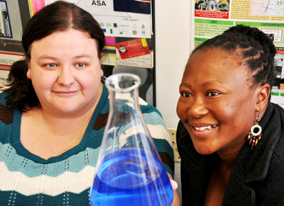
 Ph.D students, Chantel Swart and Ntsoaki Leeuw


Scientists at the University of the Free State (UFS) made an important breakthrough in the use of nanotechnology in medical and biological research. The UFS team’s research has been accepted for publication by the internationally accredited Canadian Journal of Microbiology.

The UFS study dissected yeast cells exposed to over-used cooking oil by peeling microscopically thin layers off the yeast cells through the use of nanotechnology.

The yeast cells were enlarged thousands of times to study what was going on inside the cells, whilst at the same time establishing the chemical elements the cells are composed of. This was done by making microscopically small surgical incisions into the cell walls.

This groundbreaking research opens up a host of new uses for nanotechnology, as it was the first study ever in which biological cells were surgically manipulated and at the same time elemental analysis performed through nanotechnology. According to Prof. Lodewyk Kock, head of the Division Lipid Biotechnology at the UFS, the study has far reaching implications for biological and medical research.

The research was the result of collaboration between the Department of Microbial, Biochemical and Food Biotechnology, the Department of Physics (under the leadership of Prof. Hendrik Swart) and the Centre for Microscopy (under the leadership of Prof.Pieter van Wyk).

Two Ph.D. students, Chantel Swart and Ntsoaki Leeuw, overseen by professors Kock and Van Wyk, managed to successfully prepare yeast that was exposed to over-used cooking oil (used for deep frying of food) for this first ever method of nanotechnological research.

According to Prof. Kock, a single yeast cell is approximately 5 micrometres long. “A micrometre is one millionth of a metre – in laymen’s terms, even less than the diameter of a single hair – and completely invisible to the human eye.”

Through the use of nanotechnology, the chemical composition of the surface of the yeast cells could be established by making a surgical incision into the surface. The cells could be peeled off in layers of approximately three (3) nanometres at a time to establish the effect of the oil on the yeast cell’s composition. A nanometre is one thousandth of a micrometre.

Each cell was enlarged by between 40 000 and 50 000 times. This was done by using the Department of Physics’ PHI700 Scanning Auger Nanoprobe linked to a Scanning Electron Microscope and Argon-etching. Under the guidance of Prof. Swart, Mss. Swart en Leeuw could dissect the surfaces of yeast cells exposed to over-used cooking oil. 

The study noted wart like outgrowths - some only a few nanometres in diameter – on the cell surfaces. Research concluded that these outgrowths were caused by the oil. The exposure to the oil also drastically hampered the growth of the yeast cells. (See figure 1)  

Researchers worldwide have warned about the over-usage of cooking oil for deep frying of food, as it can be linked to the cause of diseases like cancer. The over-usage of cooking oil in the preparation of food is therefore strictly regulated by laws worldwide.

The UFS-research doesn’t only show that over-used cooking oil is harmful to micro-organisms like yeast, but also suggests how nanotechnology can be used in biological and medical research on, amongst others, cancer cells.


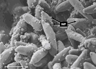
Figure 1. Yeast cells exposed to over-used cooking oil. Wart like protuberances/ outgrowths (WP) is clearly visible on the surfaces of the elongated yeast cells. With the use of nanotechnology, it is possible to peel off the warts – some with a diameter of only a few nanometres – in layers only a few nanometres thick. At the same time, the 3D-structure of the warts as well as its chemical composition can be established.  

Media Release
Issued by: Mangaliso Radebe
Assistant Director: Media Liaison
Tel: 051 401 2828
Cell: 078 460 3320
E-mail: radebemt@ufs.ac.za  
18 August 2010
 

We use cookies to make interactions with our websites and services easy and meaningful. To better understand how they are used, read more about the UFS cookie policy. By continuing to use this site you are giving us your consent to do this.

Accept