Did you know that one can photograph a plant so clearly from a distance that it is likely to detect the health of its leaves? Or can you contemplate the possibility of taking a photograph of three similar-looking rocks, being able to distinguish them
from each other due to spectral properties associated with their internal mineral content?
This and other drone-based geological imaging are made possible by the Merensky group for Aerial Geological Image Classification (MAGIC) at the University of the Free State (UFS).
Recently (24 March 2022), the President of the
Hans Merensky Foundation (HMF),
Dr Khotso Mokhele, signed a R11 million five-year research grant agreement with the UFS. Merensky research projects are currently limited to three South African universities – Stellenbosch University
(for forestry research), the University of Pretoria (for avocados), and now the UFS.
Demonstration and application
Dr Mokhele, who is also the former Chancellor of the UFS, states: “When I walked into the MAGIC Lab, I knew that something special was going to happen here. What we are launching today will become a world-class and world-leading facility.”
According to him, Dr Hans Merensky, whose legacy is facilitated by the foundation, was one of the most influential geologists in South Africa. He discovered, among others, deposits of gold, platinum, diamonds, phosphates, and vermiculite. After several
decades of operating in the geological sciences and with his knowledge of soil health, Merensky became a conservationist of note and played a key role in the establishment of the country’s agricultural practices.
“The main objective of the Hans Merensky Foundation is to promote and assist in the development of the resources of South Africa and neighbouring territories – particularly such natural resources as soil, water, flora, and fauna – and
to promote the health and welfare of the inhabitants; more specifically, through research, experimentation, and demonstration and through the correlation and application of scientific knowledge.”
“You have to take the knowledge and translate it into demonstration of what that knowledge can do, and then apply it,” says Dr Mokhele.
The grant is also evidence of Dr Merensky’s generosity. He is well quoted saying: “This country has given to me so much, that I am only too happy to be allowed to help it to develop in some way, and I am grateful to be able to give back to
it a fraction of what it has given to me.”
Next generation of scientists
The Rector and Vice-Chancellor of the UFS,
Prof Francis Petersen, said at the
signing ceremony that this is an exciting project. “For us as a university, research and the development of the next generation of scientists are critical. This is part of our mandate. This project is one of those catalysts for the development
of what the mandate is all about – research output, capacity building, and impact through our students and our research in a broader society.”
He thanked Dr Mokhele for facilitating the project and introducing the UFS to the Hans Merensky Foundation, having the confidence that the university has the capacity to deliver.
“For Dr Mokhele, it is all about the science. To a certain extent, his driving force was to use science to make a difference in the lives of people,” says Prof Petersen.
He continues: “The institution is highly committed to this project and will make sure that it receives the maximum support to make it a success.”
The grant has been allocated to MAGIC and funds will be used to support research programmes. “This includes student bursaries, staff salaries, capital expenditure acquisitions such as high-performance computers, as well as the drones that the project
makes use of,” states Dr Martin Clark, Lecturer in the
Department of Geology.
According to him, the group aims to develop drone-based geological imaging in South Africa, with specific attention to mineral and groundwater exploration endeavours.
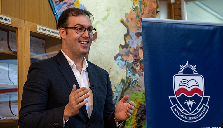
(Dr Martin Clark, principal investigator of MAGIC, says what makes him excited about this project is how the research impacts
society. Photo: Sonia Small)
Impacting society
“What makes me excited about this project is how the research impacts society. This includes developing geological imaging capacity in South African geologists with a 4IR skillset, ensuring that they remain competitive in a global market,”
says Dr Clark.
He is also of the opinion that many industries will be able to see for themselves how this technology can improve their businesses. “Drone-based geological imaging can be quicker, cheaper, and safer for collecting much of the initial information
that informs more expensive exploration processes, such as drilling. Additionally, it is non-invasive, and has little to no impact on the environment during data collection. Drones can also, in terms of safety, collect data from unstable rock walls
– historically, geologists would have to take those measurements themselves, with rock falls resulting in a significant number of deaths every year.”
Recent research
Dr Clark says drone-based imaging has supported
research initiatives in the Vredefort Dome. “Using drone-collected high-resolution images of meteorite impact
melt rocks, along with field observations of how much and where foreign rock components were contained within (clasts), we could make a case for turbulent flow in the migration of impact melt material within the deep crust.”
He adds that three papers are currently underway, each predicated on drone imagery that enables new insights into geological processes or the ability to digitally translate geological information inside and outside the classroom.
The growing research group, with Dr Clark as the principal investigator, consists of one PhD student, two master’s students, and two honours students, with several postdoctoral research fellows to follow soon.
The difference
Although several universities in the country have started using drones, the UFS has significant support to grow drone applications. With assets such as the high-performance computing cluster, very large drone-borne datasets can be resolved in record time.
“The UFS also has a wealth of world-class researchers focused on topics such as farming and environmental management, who will be able to benefit from the drone infrastructure being established on campus. We are aiming to be the go-to geological
drone imaging group in South Africa,” he says.
About mineral and groundwater exploration
Dr Martin Clark explains that drones can carry several types of cameras, from regular photographic cameras that capture photos as we know it, to thermal cameras showing differences in hot and cold bodies, to spectral cameras capturing beyond what
our eyes can see into other portions of the electromagnetic spectrum. The MAGIC group mainly uses regular and spectral cameras for their applications.
He says with mineral exploration, a high-resolution understanding of the geometry of rock bodies enables us to better identify where more costly mineral exploration techniques (e.g., drilling) should go. This process allows for a better understanding
of how geological areas have developed from a structural perspective – in essence, from where and how rocks have been displaced and deformed, and by association, the mineral deposits contained within.
“In terms of groundwater exploration, regular cameras are used to understand where rocks are fractured, where specific groundwater-influencing lithological bodies are located, and how they are orientated.”
Dr Clark continues: “With spectral cameras, we can perceive the level of access that surface plants have to water resources. It is also possible to spot the distribution of plant types associated with, or strongly dependent on, available near-surface
groundwater resources. By using spectral data, which was historically collected from satellites, we can understand how areas of land have been affected by growing, shrinking, or shifting underground bodies of water.”