The Annual Final-Year Student Exhibition of the
Department of Fine Arts at the University of the Free State is underway.
The exhibition, which is on display in the Johannes Stegmann Gallery on the ground floor of the UFS Sasol Library on the UFS Bloemfontein Campus, will be open for viewing until 24 March 2023. The exhibition event is scheduled for 1 March 2023 at 18:00.
Angela de Jesus, Curator of the UFS Art Gallery, says the exhibition comprises artworks made by the final-year Fine Arts students in the Department of Fine Arts. “Each student works with a particular research topic or theme of their choosing.”
This year’s exhibition features the work of Courtney Micaela Parsons, Megan Johns, PK Mohanoe, and Natley Bernardo.
Craft as art
Parsons, who is interested in the notion of craft as art, in particular transgenerational and traditional female crafts, has transformed a found bed into an art object with her work Stitching generational divides.
“The artwork was inspired by a copper bed that belonged to three generations of women in my family. In the artwork, I explore the relationships between these generations and the bonds formed, or the lack thereof, through traditional female teachings,” she says.
The artist feels that her artwork speaks to a wish she has to forge closer ties with her maternal line and nostalgia for moments not had. “The skill of crochet is symbolic of the skills that would have been taught by older generations, which are longed for in my own life.”
Constructing art
About his works on display, named Shoot our shot, Heavy labour, Artist on site, and Artistic labour, Mohanoe says that one of the main aims of his art is to address issues regarding contentious everyday working environments and the challenges that many workers have to face in South Africa today. These include inadequate resources, a lack of occupational health and safety measures, and being poorly paid.
“I am interested in this because I personally had to overcome many challenges in having to work part time while studying full time.”
“I explore this theme and compare it to the labour involved in artmaking and the creation process,” he adds.
Mohanoe explains that his art consists of materials that can be both building and art materials, such as wooden doors, stones, and metal. “I layer, construct, deconstruct, paint, carve, and destroy these materials and document my creative process to emphasise the intense labour that is often hidden behind a final work of art,” he says, adding that he hopes to evoke empathy and encourage appreciation towards this labour by the artist, the worker, and the labouring artist.
Visiting the exhibition, visitors can also look forward to Mohanoe’s Mosebetsi o Phethahetse (image below).
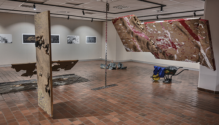
Mohanoe’s Mosebetsi o Phethahetse
Mixed media on three deconstructed doors,
metal chain, artists conti suit, wheelbarrow,
boots, stones, bricks & drop sheet
Dimensions vary
Water and the subconscious
Barnardo, who grew up in the Western Cape, was surrounded by water in her early years and longed for it after moving to the Free State.
“I experienced a sense of loss, which initiated my need to return to and surround myself with water. The water is a place of nostalgia for me, where I can experience an overflow of memories and thoughts, because I relate to its fluidity and plurality,” explains Barnardo, who has on display works named Changing mid-stream, Submerged subconscious I, II, and III, Translucent memory, and Fragmentary waters I, II.
She says that the transparent quality of water becomes the vehicle that purifies and clarifies her understanding of her own subconscious mind – a place of reminiscence that allows her to cross the threshold into the subconscious and brings hidden things to the surface.
Also on display is a video installation, named Liminal Spaces between water and the subconscious. Barnardo says she blindly takes video footage underwater, which allows her to discover and rediscover things that were hidden from view. “In doing so, I allude to the latency of images within the subconscious mind,” she says.
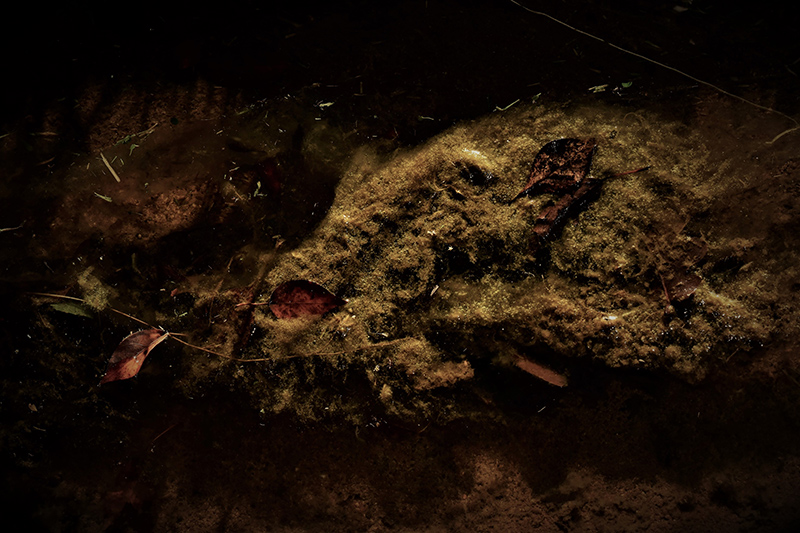
Natley Barnardo,
Submerged
subconscious I,
digital photography, 46 x 69.5cm
In her art, Johns is interested in investigating how childhood trauma and abandonment can affect adult life and how it can be the source of obsessive melancholia.
Melancholia, Johns believes, is compared to the constant reopening of an old wound. “In my artwork, the artistic practice of drawing, scribbling, or scratching represents the ‘scratching open of old wounds’. The process of scratching or marring a surface is similar to how scratching a wound mars the skin. These scratches also speak of the long-lasting damage that trauma can bring to memory. I use drawings which are reminiscent of childhood mark-making and scribbling, combining them with old family photographs and objects associated with childhood play,” describes Johns.
She has on display digital drawings and collages on perspex, including Harmonious union, Mother and daughter, and Self-portrait.
There are also two collections of this artist on display, named Puzzle, and Abandoned objects series.
• The exhibition can also be viewed online here